Q&A: Are you getting enough riboflavin (vitamin B2) for optimal energy and health?
Riboflavin (vitamin B₂) is essential for energy production, redox balance, and iron metabolism. Found in dairy, meat and fortified foods, it supports multiple metabolic pathways. Although deficiency is uncommon, suboptimal intake may impact fatigue, anaemia and chronic disease risk.


Introduction 📖
Riboflavin (vitamin B₂) was originally identified as vitamin G before being isolated and chemically characterised as 7,8-dimethyl-10-ribitylisoalloxazine (three fused six-membered rings, with a sugar alcohol side chain attached to the central ring). The name “riboflavin” reflects both its structure and appearance—“ribose” denoting its sugar moiety and flavus, the Latin word for yellow, describing its characteristic colour. It was subsequently reclassified as vitamin B₂ to reflect its position within the B-vitamin complex.
Riboflavin (vitamin B₂) is often overlooked in discussions of micronutrient health, yet it plays a central role in energy metabolism, redox balance and the function of several other vitamins. I explore the biochemical functions, dietary sources, clinical relevance and emerging therapeutic roles of riboflavin, including its potential in personalised nutrition.
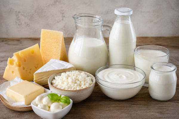
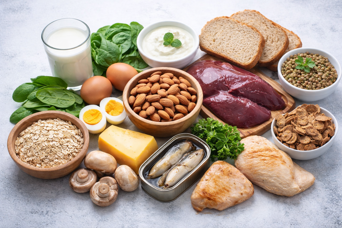
Food sources 🥗
Riboflavin is an essential water-soluble vitamin that must be obtained from dietary sources (exogenous) because the body cannot synthesise it. Riboflavin may be synthesised by lactic acid bacteria consumed in foods and by commensal gut microorganism that lives in or on the human body without causing harm, including certain Bifidobacterium species but amounts are too small to contribute to needs (1).
Riboflavin is widely distributed in foods, with particularly rich sources including liver, meat, dairy products and cereals that are either enriched or fortified. Most fruits and vegetables provide relatively small amounts, while whole grains contain modestly higher levels.
Riboflavin is also used as a food colouring agent, appearing as a yellow–orange crystalline powder and is designated as E101 in Europe for use as a food additive (https://www.food.gov.uk/business-guidance/approved-additives-and-e-numbers#colours).
Table: Riboflavin (vitamin B₂) food sources (South African context)
|
Animal
Sources (SAFOODS values; per 100 g unless stated) |
Riboflavin
(mg) |
Plant
Sources (SAFOODS values; per 100 g unless stated) |
Riboflavin
(mg) |
|
Beef liver
(cooked) |
~2.8–3.2 |
Almonds |
~0.9–1.1 |
|
Chicken
liver (cooked) |
~2.0–2.5 |
Mushrooms
(brown/white) |
~0.4–0.5 |
|
Milk, full
cream (250 ml) |
~0.4–0.5 |
Spinach /
imifino (cooked leafy greens) |
~0.2–0.3 |
|
Maas
(amasi), fermented milk (250 ml) |
~0.3–0.4 |
Fortified
maize meal (pap) |
~0.3–0.6* |
|
Yogurt
(plain, 250 ml) |
~0.4–0.5 |
Fortified
breakfast cereals |
~0.5–1.5+* |
|
Eggs (2
large) |
~0.4–0.5 |
Cooked dry
beans (e.g., sugar beans) |
~0.1–0.2 |
|
Cheddar
cheese |
~0.3–0.4 |
Lentils
(cooked) |
~0.1–0.2 |
|
Beef (lean,
cooked) |
~0.2–0.3 |
Whole wheat
bread |
~0.1–0.2* |
|
Chicken
(cooked) |
~0.1–0.2 |
Peanuts |
~0.1–0.2 |
|
Pilchards
(canned) |
~0.2–0.3 |
Avocado |
~0.1–0.2 |
Riboflavin is relatively stable during cooking but is highly sensitive to light exposure, which can lead to significant losses.
🔎 Practical tips to preserve riboflavin:
- Limit light exposure: Store milk and dairy products in opaque containers and avoid prolonged exposure to sunlight or fluorescent light.
- Avoid unnecessary soaking: As a water-soluble vitamin, riboflavin can leach into soaking or cooking water.
- Use minimal cooking water: Steaming or microwaving vegetables preserves more riboflavin than boiling.
- Retain cooking liquids where possible: For example, use broth from cooked legumes or vegetables in soups or sauces.
- Store foods properly: Keep riboflavin-rich foods in cool, dark conditions to minimise degradation.
- Consume fresh when possible: Prolonged storage, especially under light, can reduce riboflavin content.
Supplement forms
Below is a structured comparison of the main supplemental forms of riboflavin (vitamin B₂) encountered in clinical nutrition, pharmacology, and commercial products.
Table: Riboflavin supplemental forms
|
Form |
Chemical name |
Active / Coenzyme form |
Bioavailability & absorption |
Typical uses |
Notes / Considerations |
|
Riboflavin |
Riboflavin |
No (precursor) |
Good absorption via active transport (saturable at
~25–30 mg/dose) |
General supplementation, deficiency prevention |
Most common and cost-effective form |
|
Riboflavin-5′-phosphate (R5P) |
Flavin mononucleotide (FMN) |
Yes (coenzyme form) |
Requires dephosphorylation before absorption;
similar bioavailability to riboflavin |
Clinical/therapeutic use (e.g., migraine,
mitochondrial support) |
Marketed as “activated” but not necessarily superior
in healthy individuals |
|
Flavin adenine dinucleotide (FAD) |
FAD |
Yes (coenzyme form) |
Hydrolysed to FMN → riboflavin prior to absorption |
Rare in supplements; mainly intracellular cofactor |
Not typically used in oral supplements |
|
Riboflavin sodium phosphate |
Sodium riboflavin-5′-phosphate |
Yes (FMN salt form) |
Water-soluble; used in parenteral formulations |
Injectable preparations, fortification |
Improved solubility vs free riboflavin |
|
Riboflavin tetrabutyrate |
Riboflavin ester |
No (derivative) |
Lipid-soluble; slower release and absorption |
Experimental / niche formulations |
May have improved tissue retention; limited clinical
use |
Absorption, transport, storage and excretion
In the stomach, hydrochloric acid (HCl) releases riboflavin from its bound forms in food and supplements. Approximately 60–65% of free riboflavin is absorbed, primarily in the small intestine via active transport at low concentrations and facilitated diffusion at higher concentrations (2). Its bioavailability increases when physiological demand is elevated, and riboflavin from animal-based foods is generally more readily absorbed than from plant sources (3). Riboflavin in plant foods may be less accessible due to binding within plant matrices, whereas in animal foods it is more readily released and absorbed (4). In the bloodstream, riboflavin is transported bound to plasma proteins. It is converted to its coenzyme forms—flavin mononucleotide (FMN) and flavin adenine dinucleotide (FAD)—in most tissues, with particularly active conversion occurring in the small intestine, liver, heart and kidneys. Only small amounts of riboflavin are stored in the body, mainly in the liver, kidneys and heart, and excess intake is excreted in the urine.
Functions ⚙️ 🔬
Riboflavin serves as the precursor to two biologically active coenzymes, flavin mononucleotide (FMN) and flavin adenine dinucleotide (FAD), as noted above. These coenzymes—collectively referred to as flavins—play central roles in cellular redox reactions, functioning as both electron acceptors and donors in energy metabolism.¹ FAD represents the oxidised form of the coenzyme; upon reduction (gain of two hydrogen atoms, equivalent to two protons and two electrons), it is converted to FADH₂.
During the tricarboxylic acid (TCA) cycle, substrates undergo dehydrogenation reactions that release hydrogen atoms and electrons. These reducing equivalents are captured by the coenzymes nicotinamide adenine dinucleotide (NAD⁺; oxidised form) and flavin adenine dinucleotide (FAD), forming NADH and FADH₂, respectively.
Within the electron transport chain (ETC), Complex I (NADH dehydrogenase) contains riboflavin-derived FMN, which accepts electrons from NADH and transfers them to ubiquinone, a process coupled to proton pumping across the inner mitochondrial membrane. In contrast, Complex II (succinate dehydrogenase)—which also functions in the TCA cycle—contains riboflavin-derived FAD as a tightly bound prosthetic group. Here, FAD accepts electrons during the oxidation of succinate to fumarate and transfers them directly to ubiquinone without contributing to proton translocation.
The electrons are subsequently passed along the ETC, ultimately reducing oxygen to water. This electron flow drives the generation of a proton gradient that powers adenosine triphosphate (ATP) synthesis, with electrons entering via FADH₂ yielding approximately 1.5 ATP per molecule. Riboflavin coenzymes are involved in many reactions in several metabolic pathways (see the table below).
Table: Functions of riboflavin
|
Functional
domain |
Role
of FMN/FAD |
Clinical
/ Physiological significance |
Key
references |
|
Energy
production |
Electron
carriers in the ETC (Complex I & II) |
Essential
for ATP generation; deficiency may contribute to fatigue and impaired
mitochondrial function |
(5, 6) |
|
Macronutrient
metabolism |
Cofactors
in oxidation–reduction reactions in carbohydrate, fat (in fatty acid
breakdown i.e. beta-oxidation to acetyl-CoA, the enzyme fatty acyl
dehydrogenase requires FAD) and protein metabolism |
Supports
energy release from macronutrients; relevant in metabolic efficiency |
(5, 6) |
|
Cytochrome
P450 system |
Required
for flavoprotein enzymes involved in detoxification |
Supports
hepatic detoxification of drugs and xenobiotics |
(6) |
|
Niacin
(vitamin B3) synthesis |
FAD
is required for conversion of tryptophan → niacin |
Influences
NAD⁺/NADP⁺ synthesis and energy/redox metabolism |
(5) |
|
Vitamin
activation (B6, folate, B12); indirect homocysteine regulation |
Supports
enzymatic activation of pyridoxine, folate and cobalamin ·
Formation of the active vitamin B6
coenzyme (pyridoxal phosphate requires FMN ·
FAD is needed for folate metabolite
5-methyl-tetrahydrofolate synthesis |
Critical
for one-carbon metabolism, DNA synthesis, and homocysteine regulation |
(5, 7, 8) |
|
Antioxidant
defence |
Cofactor
for glutathione reductase (FAD-dependent) → regeneration of reduced
glutathione (GSH) |
Maintains
redox balance and protects against oxidative stress |
(5, 6) |
|
Redox
reactions (general) |
Central
role in oxidation–reduction reactions across metabolic pathways |
Fundamental
for cellular homeostasis and metabolic integration |
(6) |
|
Iron
metabolism and haem synthesis |
Cofactor
for flavoproteins involved in iron absorption, mobilisation, and haem
synthesis • Supports iron metabolism and
erythropoiesis |
Supports
iron utilisation and haemoglobin synthesis; deficiency may contribute to
anaemia |
(6) |
ATP, adenosine triphosphate; ETC, electron transport chain
Riboflavin (vitamin B₂) supports erythropoiesis primarily through its role as a precursor of the coenzymes flavin adenine dinucleotide (FAD) and flavin mononucleotide (FMN), which are essential for redox reactions in cellular metabolism. These flavins are required for iron metabolism, including the reduction of ferric (Fe³⁺) to ferrous (Fe²⁺) iron, facilitating its absorption, transport and incorporation into haemoglobin. Riboflavin also supports the mobilisation of iron from storage sites, thereby influencing iron availability for erythrocyte synthesis. In addition, it contributes to the maintenance of glutathione in its reduced form via the FAD-dependent enzyme glutathione reductase, protecting developing erythrocytes from oxidative damage. Indirectly, riboflavin interacts with other haematopoietic nutrients, such as vitamin B₆ and folate, further supporting haem synthesis and red blood cell maturation.
Deficiency 🚫
Riboflavin deficiency (ariboflavinosis) is relatively uncommon and typically occurs alongside deficiencies of other B vitamins. This reflects both their co-occurrence in many food sources and their interdependent roles in metabolic pathways, where the availability of one B vitamin can influence the function and utilisation of others.
Ariboflavinosis manifests primarily in rapidly regenerating tissues, leading to characteristic mucocutaneous and ocular abnormalities. Angular cheilitis reflects impaired epithelial integrity at the corners of the mouth, while glossitis (magenta tongue) indicates inflammation and atrophy of the lingual papillae. Seborrheic dermatitis, particularly in the nasolabial folds, arises from disrupted skin barrier function and altered lipid metabolism. Ocular signs such as corneal vascularisation and photophobia are linked to compromised epithelial maintenance and increased oxidative stress in the eye.
Riboflavin (vitamin B2) deficiency impairs iron status through multiple mechanisms, including reduced intestinal iron absorption, disrupted mobilisation of iron from stores and impaired haem synthesis. As a result, iron cannot be effectively utilised for haemoglobin production, increasing the risk of functional iron deficiency and anaemia (normocytic anaemia) even when dietary iron intake is adequate.
Deficiency is most likely in, individuals with alcohol use disorder (due to reduced intake, absorption and hepatic utilisation), the elderly (often with inadequate dietary intake), those with poor dietary diversity and patients with chronic illness (cancer, certain forms of cardiovascular disease and diabetes (9, 10)) or malabsorption syndromes that impair nutrient uptake. Individuals who follow vegan diets or consume little dairy may be at increased risk of riboflavin inadequacy.
Additionally, certain medications can interfere with riboflavin metabolism or increase its requirements, further elevating deficiency risk. Oral contraceptives have been associated with reduced riboflavin levels (11), phenobarbital and other anticonvulsants may increase riboflavin metabolism and tricyclic antidepressants (10) can interfere with its utilisation, collectively increasing the risk of suboptimal riboflavin status.
A separate high-risk group includes individuals with riboflavin transporter deficiency, a rare genetic disorder caused by mutations in riboflavin transporter genes (SLC52A2 or SLC52A3) (12). These mutations impair the absorption and transport of riboflavin, leading to deficiency and neurological symptoms such as hearing loss, bulbar dysfunction and respiratory complications (12). Although there is no cure, early initiation of high-dose riboflavin supplementation can be life-saving and improve outcomes.
Toxicity and safety⚠️
Riboflavin (vitamin B₂) has very low toxicity, and no tolerable upper intake level has been established due to the absence of consistent evidence of adverse effects (10, 13). This is largely attributable to its water-soluble nature, limited intestinal absorption at high doses, and efficient renal excretion of excess amounts. Even at supplemental doses far exceeding physiological requirements, riboflavin is generally well tolerated, with the most notable effect being harmless flavinuria (bright yellow urine) (14). Rarely, very high doses may cause mild gastrointestinal discomfort, but clinically significant toxicity has not been demonstrated.
Clinical conditions and therapeutic roles 💊 🩺
Homocysteine, MTHFR and blood pressure ❤️
Riboflavin (vitamin B2), in its coenzyme form flavin adenine dinucleotide (FAD), is essential for the activity and stability of methylenetetrahydrofolate reductase (MTHFR), a key enzyme in folate metabolism. MTHFR converts folate into its active form, 5-methyltetrahydrofolate (5-MTHF), which is required for the remethylation of homocysteine (an amino acid associated with cardiovascular risk) to methionine, thereby supporting normal methylation processes.
FAD is also required for enzymes involved in the activation of vitamin B6 (conversion of pyridoxal to pyridoxal-5′-phosphate)(8), highlighting the broader role of riboflavin in one-carbon metabolism. Consequently, inadequate riboflavin status may impair these pathways and contribute to elevated plasma homocysteine concentrations, both directly through reduced MTHFR activity and indirectly through effects on other B vitamins (8).
The influence of riboflavin on homocysteine is supported by some epidemiological evidence (e.g., the Framingham Offspring Study (15)), although findings are inconsistent in the general population (16). Riboflavin status appears particularly relevant in individuals with the MTHFR C677T polymorphism (17). The 677T allele reduces the binding affinity of MTHFR for FAD, leading to decreased enzyme activity and higher homocysteine levels.
Moreover, riboflavin status can influence the efficiency of folate utilisation, particularly in individuals with the MTHFR C677T polymorphism, where reduced enzyme affinity for FAD leads to impaired activity (18). Evidence indicates that adequate riboflavin status enhances the metabolic response to folate and lowers homocysteine concentrations, highlighting a synergistic interaction between these nutrients. This interaction appears most pronounced in individuals with the 677TT genotype but may also be relevant at the population level, particularly in settings of suboptimal folate intake.
Beyond homocysteine metabolism, this genetic variant is also associated with elevated blood pressure and increased risk of hypertension. Notably, genome-wide studies (19) and randomised controlled trials (20) have demonstrated that riboflavin supplementation can lower blood pressure in individuals with this genotype, in some cases more effectively than antihypertensive treatment alone and randomised controlled trials have shown that riboflavin supplementation can lower blood pressure in affected individuals, in some cases more effectively than antihypertensive treatment alone. H-type hypertension is defined as the coexistence of elevated blood pressure and hyperhomocysteinaemia, representing a higher-risk cardiovascular phenotype. In an observational study I co-authored, riboflavin intake was lower in individuals with H-type hypertension compared to those with hypertension without elevated homocysteine levels (21). These findings highlight the potential for safe, targeted interventions such as riboflavin supplementation and support the broader concept of personalised nutrition and treatment based on genetic risk factors.
Cancer 🎗️🧬
Riboflavin may contribute to cancer prevention through its role as a coenzyme in cytochrome P450 systems, where it supports the detoxification of carcinogens and may help reduce DNA damage. Despite this plausible biological mechanism, evidence from human studies remains limited, and there is a notable lack of randomised controlled trials to establish causality. Current evidence is therefore largely derived from observational studies which suggest a possible protective association between riboflavin intake and certain cancers.
Table: Meta-analysis on riboflavin intake and cancer risk
|
Study design |
Key findings |
Reference |
|
Systematic review and meta-analysis of 8 observational studies (4
cohort, 4 case-control; n = 7,750 colorectal cancer cases); random-effects
model |
Higher riboflavin intake was associated with a reduced risk of
colorectal cancer (OR = 0.83; 95% CI: 0.75–0.91). Sensitivity analysis
confirmed robustness (OR = 0.86). Findings suggest an inverse association,
though causality remains uncertain |
(22) |
|
Systematic review and meta-analysis of 10 epidemiological studies (n =
12,268 breast cancer cases); pooled using random-effects model; publication
bias assessed (Egger’s test) |
Higher dietary riboflavin intake was associated with a modest
reduction in breast cancer risk (RR = 0.85; 95% CI: 0.76–0.95). Dose–response
analysis showed that each 1 mg/day increase in intake was linked to a 6%
reduction in risk (RR = 0.94; 95% CI: 0.90–0.99). No significant
heterogeneity or publication bias was detected. Overall, findings suggest a weak
inverse association, but not definitive protection. |
(23) |
CI, confidence interval; OR, odds ratio; RR, relative risk
Migraine 🧠 ⚡
A systematic review and dose–response meta-analysis of 12 clinical trials (n = 749) found that riboflavin supplementation reduces migraine frequency and duration, with greater benefits observed at higher doses up to 400 mg/day (24). The findings suggest a dose-dependent effect without a clear threshold, although limitations such as small sample sizes and study heterogeneity should be considered (24).
Corneal thinning 👁️
Riboflavin plays a clinically important role in the structural integrity of the cornea, particularly through its involvement in collagen stabilisation. In ophthalmology, riboflavin is central to corneal collagen cross-linking (CXL), a therapeutic procedure used to halt the progression of corneal thinning disorders such as keratoconus. During CXL, topical riboflavin (vitamin B₂) is applied to the cornea and activated with ultraviolet-A (UVA) light, generating reactive oxygen species that induce covalent cross-links between collagen fibres. This process increases corneal rigidity and resistance to further thinning or deformation.
Requirements 📊
Riboflavin requirements vary across the life course and according to physiological and metabolic demands. For adults, recommended intakes are approximately 1.1 mg/day for women and 1.3 mg/day for men (10), with lower requirements in children depending on age. Needs increase during pregnancy (≈1.4 mg/day) and lactation (≈1.6 mg/day) to support foetal development and milk production (10). Requirements may also be elevated in individuals with high metabolic demand, such as athletes, or during illness (because riboflavin decrease in the context of inflammation (25)) and physiological stress, where turnover and utilisation of flavin coenzymes are increased. Emerging evidence suggests that individuals with diabetes may have altered riboflavin status due to increased oxidative stress and metabolic demands (9). T allele carriers of the MTHFR C677T polymorphism (17) or mutations in riboflavin transporter genes (SLC52A2 or SLC52A3) may also need more riboflavin (11).
Assessment of status 🧪
The erythrocyte glutathione reductase activity coefficient (EGRAC) is considered the gold standard functional biomarker of riboflavin status because it reflects intracellular flavin cofactor availability (10). Glutathione reductase is a flavin adenine dinucleotide (FAD)-dependent enzyme, and its activity increases in vitro when FAD is added if riboflavin status is suboptimal. Thus, a higher EGRAC value indicates poorer riboflavin status. In contrast, plasma riboflavin concentrations are less reliable, as they are influenced by recent dietary intake and do not accurately reflect tissue stores or functional sufficiency.
Conclusion 🧾
Riboflavin is a fundamental yet frequently underappreciated nutrient that underpins key metabolic, haematological and cellular processes. Its role extends beyond energy production to include antioxidant defence, vitamin activation and interactions with genetic pathways influencing cardiovascular health and possibly cancer risk. Although deficiency is often subtle, its clinical implications—particularly for iron metabolism and redox balance—are significant. Ensuring adequate intake through a diverse diet or targeted supplementation where necessary represents a practical and evidence-based strategy to support overall health and metabolic resilience.
Reflections 🤔 💭
· Do I regularly consume riboflavin-rich foods such as dairy, eggs, meat, or fortified cereals?
· Do I frequently consume alcohol, which may impair riboflavin absorption?
· Am I taking medications (e.g., oral contraceptives, anticonvulsants, antidepressants) that may affect riboflavin status?
· Have I had signs of iron deficiency despite adequate iron intake?
· Have I ever had my nutritional status assessed, particularly if I have a chronic condition or restricted diet?
· Do I have a diagnosis of hypertension that is difficult to control despite treatment? Have I been tested for, or do I suspect, the MTHFR C677T polymorphism, which may influence my response to riboflavin? Would a personalised nutrition approach—including riboflavin optimisation—be relevant to my blood pressure management?
· Do I experience frequent or recurrent migraines, particularly those that are resistant to standard treatment? Have I ever trialled riboflavin supplementation (e.g., ~400 mg/day) under guidance, and did it affect migraine frequency or severity?
Love what you’re reading? Share the knowledge! 📨 Forward this link to a friend who might also enjoy it.
References 📚
1. Tamés H, Cuesta I, Vázquez X, López P, Ruiz L, Ruas-Madiedo P. Characterization of riboflavin-overproducing Bifidobacterium longum subsp. infantis strains selected by roseoflavin treatment. Food Microbiology. 2025;131:104799.
2. Dainty JR, Bullock NR, Hart DJ, Hewson AT, Turner R, Finglas PM, et al. Quantification of the bioavailability of riboflavin from foods by use of stable-isotope labels and kinetic modeling. The American journal of clinical nutrition. 2007;85(6):1557-64.
3. Chungchunlam SMS, Moughan PJ. Comparative bioavailability of vitamins in human foods sourced from animals and plants. Crit Rev Food Sci Nutr. 2024;64(31):11590-625.
4. Beltramo B, Urlings M, Padilla-Díaz CM, Bast A, Diliën H, de Boer A. Bioavailability of vitamins C, B2 and B9 (Folate) in nutrition and health claims: a critical appraisal. Food Production, Processing and Nutrition. 2025;7(1):55.
5. McNulty H, Pentieva K, Ward M. Causes and clinical sequelae of riboflavin deficiency. Annual Review of Nutrition. 2023;43(1):101-22.
6. Powers HJ. Riboflavin (vitamin B-2) and health. The American journal of clinical nutrition. 2003;77(6):1352-60.
7. Bailey LB, Gregory III JF. Folate metabolism and requirements. The Journal of nutrition. 1999;129(4):779-82.
8. Scott JM, editor Genetic variation of homocysteine metabolism and atherosclerosis. Nestle Nutrition Workshop Series; 2003: Philadelphia; Lippincott-Raven; 1999.
9. Cole HS, Lopez R, Cooperman JM. Riboflavin deficiency in children with diabetes mellitus. Acta diabetologia latina. 1976;13(1):25-9.
10. Berger MM, Shenkin A, Schweinlin A, Amrein K, Augsburger M, Biesalski H-K, et al. ESPEN micronutrient guideline. Clinical Nutrition. 2022;41(6):1357-424.
11. Tonkin SY. Vitamins and oral contraceptives. Vitamins In Human Biology and Medicine (1981): CRC Press; 2017. p. 29-64.
12. Jaeger B, Bosch AM. Clinical presentation and outcome of riboflavin transporter deficiency: mini review after five years of experience. Journal of inherited metabolic disease. 2016;39(4):559-64.
13. Nutrients SoURLo, Intakes SCotSEoDR, Folate iPo, Vitamins OB, Choline. Dietary reference intakes for thiamin, riboflavin, niacin, vitamin B6, folate, vitamin B12, pantothenic acid, biotin, and choline. 2000.
14. West DW, Owen EC. The urinary excretion of metabolites of riboflavine by man. British Journal of Nutrition. 1969;23(4):889-98.
15. Jacques PF, Bostom AG, Wilson PW, Rich S, Rosenberg IH, Selhub J. Determinants of plasma total homocysteine concentration in the Framingham Offspring cohort. The American journal of clinical nutrition. 2001;73(3):613-21.
16. Verhoef P, de Groot LC, editors. Dietary determinants of plasma homocysteine concentrations. Seminars in vascular medicine; 2005: Copyright© 2005 by Thieme Medical Publishers, Inc., 333 Seventh Avenue, New ….
17. Hustad S, Ueland PM, Vollset SE, Zhang Y, Bjørke-Monsen AL, Schneede J. Riboflavin as a determinant of plasma total homocysteine: effect modification by the methylenetetrahydrofolate reductase C677T polymorphism. Clinical chemistry. 2000;46(8):1065-71.
18. Moat SJ, Ashfield-Watt PA, Powers HJ, Newcombe RG, McDowell IF. Effect of riboflavin status on the homocysteine-lowering effect of folate in relation to the MTHFR (C677T) genotype. Clinical chemistry. 2003;49(2):295-302.
19. McNulty H, Strain J, Hughes CF, Ward M. Riboflavin, MTHFR genotype and blood pressure: a personalized approach to prevention and treatment of hypertension. Molecular aspects of medicine. 2017;53:2-9.
20. Wilson CP, McNulty H, Ward M, Strain J, Trouton TG, Hoeft BA, et al. Blood pressure in treated hypertensive individuals with the MTHFR 677TT genotype is responsive to intervention with riboflavin: findings of a targeted randomized trial. Hypertension (Dallas, Tex : 1979). 2013;61(6):1302-8.
21. du Plessis JP, Lammertyn L, Schutte AE, Nienaber-Rousseau C. Homocysteine, blood pressure and gene–diet interactions in relation to vascular function measures of black South Africans. Genes & Nutrition. 2024;19(1):14.
22. Liu Y, Yu Q-Y, Zhu Z-L, Tang P-Y, Li K. Vitamin B2 intake and the risk of colorectal cancer: a meta-analysis of observational studies. Asian Pacific Journal of Cancer Prevention. 2015;16(3):909-13.
23. Yu L, Tan Y, Zhu L. Dietary vitamin B2 intake and breast cancer risk: a systematic review and meta-analysis. Archives of Gynecology and Obstetrics. 2017;295(3):721-9.
24. Amini S, Heidari Z, Clark CCT, Bagherniya M. The effect of riboflavin on the mean attack frequency, severity, and duration of migraine headaches: A systematic review and dose–response meta-analysis of clinical trials. Journal of Research in Medical Sciences. 2026;31(1).
25. McMillan DC, Maguire D, Talwar D. Relationship between nutritional status and the systemic inflammatory response: micronutrients. Proceedings of the Nutrition Society. 2019;78(1):56-67.
In developing this work, the author utilised ChatGPT-4 to assist with language editing.